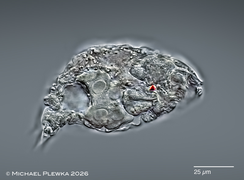
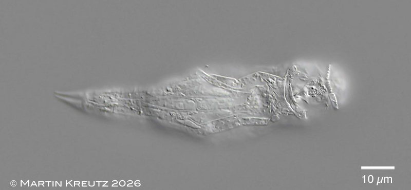
Asciaporrecta hyalina: lateral view of a slightly compressed specimen. Focal plane on the red cerebral eyespot. (1)
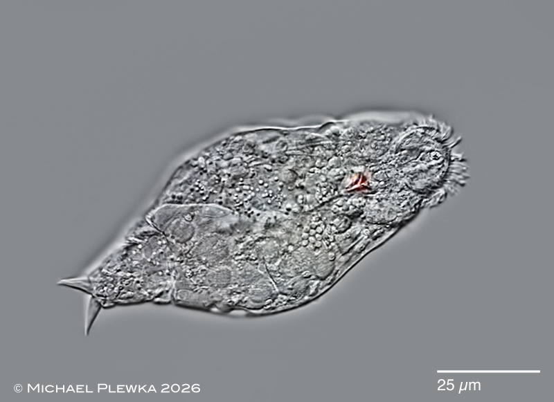
Asciaporrecta hyalina: dorsoventral view of a slightly compressed specimen. Focal plane on the oblique corona. (1)
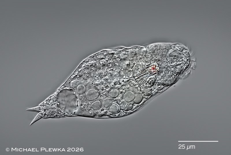
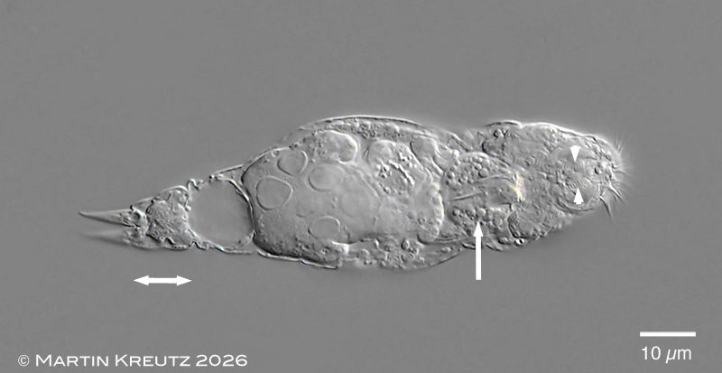
Asciaporrecta hyalina: dorsoventral view of a slightly compressed specimen. Focal plane on the mastax with granules inside. Also visible are the 8 very pronounced nuclei of the vitellarium and the rounded bladder. (1)
Asciaporrecta hyalina: two aspects of the ventral side; upper image: focal plane on the ventral ciliary field (part of); lower image: focal plane on 3 dentated structures of the corona (2)
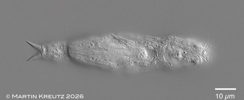
Asciaporrecta hyalina: same specimen; dorsoventral view / optical transect. The double arrow marks the foot with 1 (?or 2?) pseudosegment (in contrast to Asciaporrecta difflugicola with 5). The arrow points to the mastax with conspicuous granules. (2)
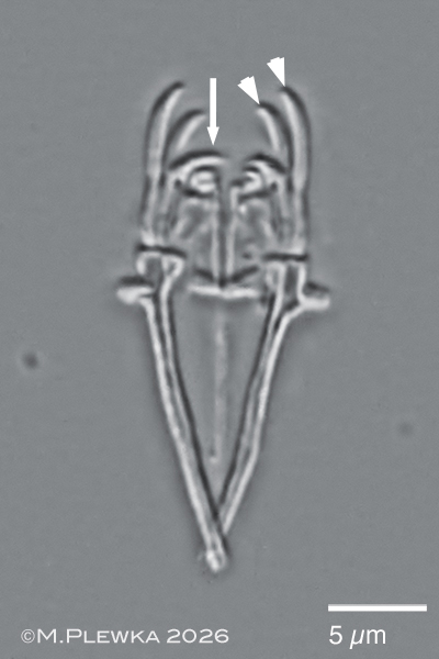
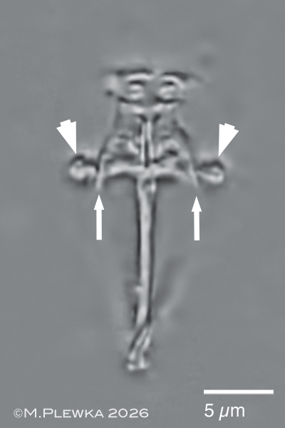
Asciaporrecta hyalina: hemiforcipate trophi of specimen from (1), dorsal view. Left: focal plane on the conspicuous unci wth 2 stout teeth (marked on the right part by arrowheads) each; subunci with curved teeth (marked on the left side by arrow). According to DeSmet only the unci/subunci participate in the grasping action, which is in contrast to Ituridae/ Dicranophoridae with pincer-shaped rami that are involved in the uptake of diet. Right: incus, focal plane on the triangular rami with pointed alulae (arrows). The round structures (marked by arrowheads) are the proximal heads of the manubria (the manubria themselves are not in focus) (1)